


The anatomical structure of the venous system of the lower extremities is characterized by great variability.Knowledge of the individual features of the structure of the venous system plays an important role in evaluating instrumental examination data and choosing the correct treatment method.
The veins of the lower extremities are divided into superficial and deep.The superficial venous system of the lower extremities begins in the venous plexuses of the toes, forming the venous network of the dorsum of the foot and the dorsal cutaneous arch of the foot.The medial and lateral marginal veins originate from it, which flow into the greater and lesser saphenous veins, respectively.The great saphenous vein is the longest vein in the body, contains 5 to 10 pairs of valves and its normal diameter is 3 to 5 mm.It originates in the lower third of the leg, in front of the medial epicondyle, and ascends into the subcutaneous tissue of the leg and thigh.In the groin region, the great saphenous vein drains into the femoral vein.Sometimes the great saphenous vein of the thigh and leg can be represented by two or even three trunks.The small saphenous vein begins in the lower third of the leg along its lateral surface.In 25% of cases, it flows into the popliteal vein in the region of the popliteal fossa.In other cases, the small saphenous vein may rise above the popliteal fossa and flow into the femoral great saphenous vein or the deep vein of the thigh.
The deep veins on the dorsum of the foot begin in the dorsal metatarsal veins of the foot, which flow into the dorsal venous arch of the foot, from where blood flows to the anterior tibial veins.At the level of the upper third of the leg, the anterior and posterior tibial veins merge to form the popliteal vein, which is located lateral and slightly posterior to the artery of the same name.In the area of the popliteal fossa, the small saphenous vein and the veins of the knee joint flow into the popliteal vein.The deep vein of the thigh usually flows into the femoral vein 6 to 8 cm below the inguinal fold.Above the inguinal ligament, this vessel receives the epigastric vein, the deep vein that surrounds the ilium, and passes into the external iliac vein, which merges with the internal iliac vein at the sacroiliac joint.The paired common iliac vein begins after the confluence of the external and internal iliac veins.The right and left common iliac veins merge to form the inferior vena cava.It is a large vessel without valves, 19 to 20 cm long and 0.2 to 0.4 cm in diameter.The inferior vena cava has parietal and visceral branches, through which blood flows from the lower extremities, lower part of the trunk, abdominal organs and small pelvis.
The perforating (communicating) veins connect the deep veins with the superficial veins.Most of them have valves located suprafascially and through which blood passes from the superficial to the deep veins.There are direct and indirect perforating veins.The direct ones directly connect the deep and superficial venous networks, the indirect ones connect indirectly, that is, they first flow into the muscular vein, which then flows into the deep vein.
The vast majority of perforating veins arise from tributaries and not from the trunk of the great saphenous vein.In 90% of patients, there is incompetence of the perforating veins on the medial aspect of the lower third of the leg.In the leg, incompetence of the perforating veins of Cockett, which connect the posterior branch of the great saphenous vein (Leonardo's vein) to the deep veins, is most often observed.In the middle and lower thirds of the thigh there are usually 2 to 4 more permanent perforating veins (Dodd, Gunter), directly connecting the trunk of the great saphenous vein with the femoral vein.In varicose transformation of the small saphenous vein, incompetent communicating veins are most frequently observed in the middle third, lower third of the leg and in the region of the lateral malleolus.
Clinical course of the disease
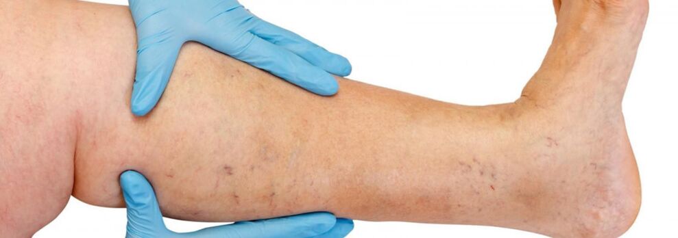
Primarily, varicose veins occur in the great saphenous vein system, less frequently in the small saphenous vein system, and begin in the tributaries of the vein trunk in the legs.The natural course of the disease in the initial phase is quite favorable;During the first 10 years or so, other than a cosmetic defect, patients may not be bothered by anything.Subsequently, if timely treatment is not carried out, complaints begin to appear of a feeling of heaviness, tiredness in the legs and swelling after physical activities (long walks, standing) or in the afternoon, especially in the hot season.Most patients complain of pain in the legs, but after detailed questioning it is possible to reveal that it is precisely a feeling of fullness, heaviness and fullness in the legs.Even with a short rest and elevated position of the limb, the severity of the sensations decreases.These symptoms characterize venous insufficiency at this stage of the disease.If we talk about pain, it is necessary to exclude other causes (arterial insufficiency of the lower extremities, acute venous thrombosis, joint pain, etc.).The subsequent progression of the disease, in addition to the increase in the number and size of dilated veins, leads to the occurrence of trophic disorders, often due to the addition of incompetent perforating veins and the occurrence of valve insufficiency of the deep veins.
In case of insufficiency of perforating veins, trophic disorders are limited to any of the surfaces of the leg (lateral, medial, posterior).Trophic disorders in the initial phase are manifested by local hyperpigmentation of the skin, followed by thickening (hardening) of the subcutaneous adipose tissue until the development of cellulite.This process ends with the formation of an ulcerative-necrotic defect, which can reach a diameter of 10 cm or more and extend deep into the fascia.The typical site of occurrence of venous trophic ulcers is the medial malleolus area, but the location of ulcers on the leg can be different and multiple.In the phase of trophic disorders, severe itching and burning occur in the affected area;Some patients develop microbial eczema.Pain in the ulcer area may not be expressed, although in some cases it is intense.At this stage of the disease, the heaviness and swelling in the legs become constant.
Diagnosis of varicose veins
It is especially difficult to diagnose the preclinical stage of varicose veins, since this patient may not have varicose veins in the legs.
In such patients, the diagnosis of varicose veins in the legs is mistakenly rejected, although there are symptoms of varicose veins, signs that the patient has relatives suffering from this disease (hereditary predisposition) and ultrasound data on initial pathological changes in the venous system.
All this can lead to failure to meet the deadlines for the optimal start of treatment, the formation of irreversible changes in the venous wall and the development of very serious and dangerous complications of varicose veins.Only when the disease is recognized at an early preclinical stage is it possible to prevent pathological changes in the venous system of the legs through minimal therapeutic effects on varicose veins.
Avoiding various types of diagnostic errors and making a correct diagnosis is possible only after a thorough examination of the patient by an experienced specialist, correct interpretation of all his complaints, a detailed analysis of the history of the disease and as much information as possible about the state of the venous system of the legs obtained using the most modern equipment (instrumental diagnostic methods).
Duplex scanning is sometimes performed to determine the exact location of perforating veins by identifying venovenous reflux in a color code.In case of valve insufficiency, your valves stop closing completely during the Valsava maneuver or compression tests.Valvular insufficiency leads to the appearance of venovenous reflux, high, through the incompetent saphenofemoral junction, and low, through the incompetent perforating veins of the leg.Using this method, it is possible to record the reverse flow of blood through the prolapsed leaflets of an incompetent valve.This is why diagnosis is multi-stage or multi-level.In a normal situation, the diagnosis is made after ultrasound diagnosis and examination by a phlebologist.However, in particularly difficult cases, the examination must be carried out in stages.
- First, a thorough examination and questioning is carried out by a phlebologist surgeon;
- if necessary, the patient is referred to additional instrumental research methods (duplex angioscanning, phleboscintigraphy, lymphoscintigraphy);
- patients with concomitant diseases (osteochondrosis, varicose eczema, lymphovenous insufficiency) receive consultation with leading consultants specializing in these diseases) or additional research methods;
- All patients requiring surgery are first consulted by the operating surgeon and, if necessary, by an anesthetist.
Treatment
Conservative treatment is mainly indicated for patients who have contraindications to surgical treatment: due to their general condition, with mild dilation of the veins causing only aesthetic disorders, or if surgical intervention is refused.Conservative treatment aims to prevent the development of the disease.In these cases, patients should be instructed to bandage the affected surface with an elastic bandage or wear elastic stockings, periodically place their legs in a horizontal position and perform special exercises for the foot and leg (flexion and extension in the ankle and knee joints) to activate the muscular-venous pump.Elastic compression accelerates and increases blood flow in the deep veins of the thigh, reduces the amount of blood in the saphenous veins, prevents the formation of edema, improves microcirculation and helps to normalize metabolic processes in tissues.Dressing should start in the morning, before getting out of bed.The bandage is applied with light tension from the toes to the thigh, with obligatory grip on the heel and ankle joint.Each subsequent round of dressing should overlap the previous one by half.It is recommended to use certified medical knitwear with individual selection of the degree of compression (from 1 to 4).Patients should wear comfortable shoes with hard soles and low heels, avoid standing for a long time, perform heavy physical work and work in hot and humid areas.If, due to the nature of the work activity, the patient has to sit for a long time, the legs must be placed in an elevated position, placing a special support of the required height under the feet.It is advisable to walk a little every 1-1.5 hours or stand on your toes 10-15 times.The resulting contractions of the calf muscles improve blood circulation and increase venous flow.While sleeping, your legs need to be placed in an elevated position.
Patients are advised to limit water and salt intake, normalize body weight and periodically take diuretics and medications that improve venous tone.According to indications, drugs are prescribed that improve microcirculation in tissues.For treatment, the use of non-steroidal anti-inflammatory drugs is recommended.
Physiotherapy plays a significant role in preventing varicose veins.For uncomplicated forms, water procedures are useful, especially swimming, hot foot baths (no higher than 35°) with a 5-10% solution of table salt.
Compressive sclerotherapy
The indications for injection therapy (sclerotherapy) for varicose veins are still being debated.The method consists of the introduction of a sclerosing agent into the dilated vein, its subsequent compression, desolation and sclerosis.Modern medicines used for these purposes are quite safe, that is, they do not cause necrosis of the skin or subcutaneous tissue when administered extravasally.Some experts use sclerotherapy for almost all forms of varicose veins, while others reject the method completely.Most likely, the truth is somewhere in between, and it makes sense for young women with the initial stages of the disease to use the injection method of treatment.The only thing is that they should be warned about the possibility of relapse (greater than with surgical intervention), the need to constantly wear a fixing compression bandage for a long period (up to 3-6 weeks) and the likelihood that several sessions may be required for complete sclerosis of the veins.
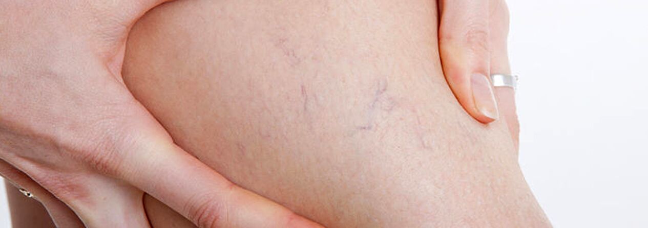
The group of patients with varicose veins should include patients with telangiectasias (“small spider veins”) and dilation of the mesh of the small saphenous veins, since the causes of the development of these diseases are identical.In this case, along with sclerotherapy, you can alsopercutaneous laser coagulation, but only after excluding damage to deep and perforating veins.
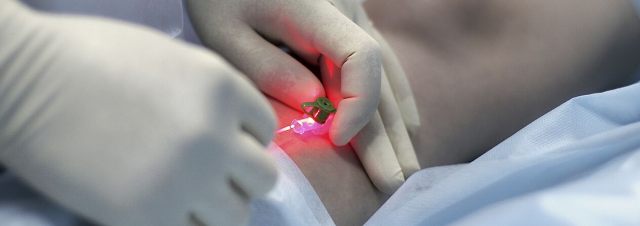
Percutaneous laser coagulation (PLC)
This is a method based on the principle of selective photocoagulation (photothermolysis), based on the different absorption of laser energy by different substances in the body.A special feature of the method is the contactless nature of this technology.The focusing head focuses energy on a blood vessel in the skin.The hemoglobin in the vessel selectively absorbs laser beams of a certain wavelength.Under the action of a laser, the destruction of the endothelium in the lumen of the vessel occurs, which leads to gluing of the vessel walls.
The effectiveness of PLK directly depends on the penetration depth of the laser radiation: the deeper the vessel, the longer the wavelength must be, therefore PLK has very limited indications.For vessels with a diameter of more than 1.0-1.5 mm, microsclerotherapy is more effective.Considering the extensive and branched distribution of spider veins on the legs and the variable diameter of the vessels, a combined treatment method is currently actively used: at the first stage, sclerotherapy of veins with a diameter of more than 0.5 mm is carried out, then a laser is used to remove the remaining “stars” of smaller diameter.
The procedure is practically painless and safe (skin cooling and anesthetics are not used), since the light from the device belongs to the visible part of the spectrum, and the wavelength of the light is designed so that the water in the tissues does not boil and the patient does not receive burns.For patients with high sensitivity to pain, preliminary application of a cream with a local anesthetic effect is recommended.Erythema and swelling subside within 1-2 days.After the course, for about two weeks, some patients may experience darkening or lightening of the treated area of the skin, which then disappears.In people with fair skin, the changes are almost imperceptible, but in patients with dark skin or a strong tan, the risk of this temporary pigmentation is quite high.
The number of procedures depends on the complexity of the case - the blood vessels are at different depths, the lesions can be small or occupy a fairly large skin surface, but usually no more than four laser therapy sessions are required (5-10 minutes each).The maximum result in such a short time is achieved due to the unique “square” shape of the device's light pulse;increases its effectiveness compared to other devices, also reducing the possibility of side effects after the procedure.
Surgical treatment
Surgery is the only radical treatment method for patients with varicose veins in the lower extremities.The purpose of the operation is to eliminate pathogenetic mechanisms (venovenous reflux).This is achieved by removing the main trunks of the great and small saphenous vein and ligating the incompetent communicating veins.
The surgical treatment of varicose veins has a centuries-old history.Previously, and many surgeons still do, large incisions along the varicose veins and general or spinal anesthesia were used.The remains after this “mini-phlebectomy” remain a lifelong reminder of the surgery.The first vein operations (according to Schade, according to Madelung) were so traumatic that the damage from them exceeded the damage from varicose veins.
In 1908, the American surgeon Babcock created a method of subcutaneous vein extraction using a rigid metal probe with an olive.In an improved form, this surgical method for removing varicose veins is still used in many public hospitals.Varicose veins are removed through separate incisions as suggested by surgeon Narat.Thus, classic phlebectomy is called the Babcock-Narat method.Phlebectomy according to Babcock-Narat has disadvantages - large scars after surgery and decreased skin sensitivity.Working capacity decreases for 2 to 4 weeks, which makes it difficult for patients to agree to surgical treatment of varicose veins.
Phlebologists have developed a unique technology for treating varicose veins in one day.Complex cases are operated withcombined technology.The main large varicose trunks are removed by inversion stripping, which involves minimal intervention through mini-incisions (2 to 7 mm) in the skin, which leave virtually no scars.The use of a minimally invasive technique involves minimal tissue trauma.The result of this operation is the elimination of varicose veins with excellent aesthetic results.Combined surgical treatment is performed under total intravenous anesthesia or spinal anesthesia, with a maximum hospitalization period of up to 1 day.
Surgical treatment includes:
- Crossectomy - crossing of the place where the trunk of the great saphenous vein enters the deep venous system;
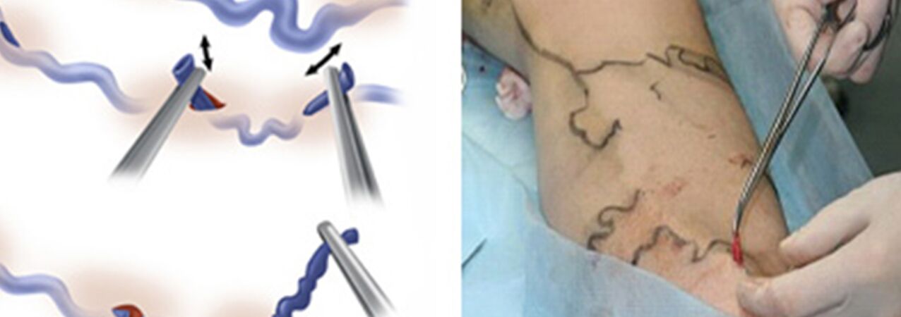
- Stripping is the removal of a varicose vein fragment.Only the varicose vein is removed, not all of it (as in the classic version).
In truthminiphlebectomyreplaced the Narat technique for removing varicose tributaries from the main veins.Previously, skin incisions measuring 1-2 to 5-6 cm were made along the course of the varicose veins, where the veins were isolated and removed.The desire to improve the cosmetic result of the intervention and to be able to remove veins not through traditional incisions, but through mini-incisions (punctures), forced doctors to develop tools that allowed them to do almost the same thing through a minimal skin defect.This is how sets of phlebectomy “hooks” of different sizes and configurations and special spatulas appeared.And instead of an ordinary scalpel, scalpels with a very narrow blade or needles with a rather large diameter for piercing the skin began to be used (for example, a needle used to take venous blood for analysis with a diameter of 18G).Ideally, the mark from a puncture with this needle becomes practically invisible after some time.
Some forms of varicose veins are treated on an outpatient basis under local anesthesia.The minimal trauma during miniphlebectomy, as well as the low risk of intervention, allow this operation to be performed in a day hospital.After minimal observation in the clinic after surgery, the patient may be sent home on their own.Postoperatively, an active lifestyle is maintained and active walking is encouraged.Temporary incapacity for work usually lasts no more than 7 days, then it is possible to start work.
When is microphlebectomy used?
- When the diameter of the varicose trunks of the great or small saphenous vein is greater than 10 mm;
- After suffering thrombophlebitis of the main subcutaneous trunks;
- After recanalization of the trunks after other types of treatment (EVLT, sclerotherapy);
- Removal of very large individual varicose veins.
It can be an independent operation or be part of a combined varicose vein treatment, combined with laser vein treatment and sclerotherapy.The tactics of use are determined individually, always taking into account the results of duplex ultrasound of the patient's venous system.Microphlebetomy is used to remove veins from various locations that have changed for a variety of reasons, including on the face.Professor Varadi of Frankfurt developed his convenient instruments and formulated the basic postulates of modern microphlebectomy.The Varadi phlebectomy method provides excellent cosmetic results without pain or hospitalization.It is very meticulous work, almost like jewelry.
After vein surgery
The postoperative period after the usual “classic” phlebectomy is quite painful.Sometimes large bruises are a concern and swelling occurs.Wound healing depends on the phlebologist's surgical technique;sometimes there is lymph leakage and prolonged formation of visible scars;Often, after a major phlebectomy, a loss of sensation remains in the heel area.
On the other hand, after miniphlebectomy, the wounds do not require suturing, as they are just punctures, there is no pain and in practice no damage to the cutaneous nerves was observed.However, such phlebectomy results are achieved only by very experienced phlebologists.